
Floor Of The Cranium Labeled Viewfloor.co
This article will describe the anatomy from the inferior view of the skull base. We will explore the many foramina and projections that enable arteries and nerves to both enter and leave the skull.

Skull diagram, superior view of floor of cranium with labels Axial
Skull superior view by khatler11 14,505 plays 14 questions ~40 sec English 14p 18 4.57 (you: not rated) Tries Unlimited [?] Last Played November 27, 2023 - 01:53 am There is a printable worksheet available for download here so you can take the quiz with pen and paper. Remaining 0 Correct 0 Wrong 0 Press play! 0% 0:00.0 Other Games of Interest

The Skull Anatomy and Physiology I
Figure 1. Parts of the Skull. The skull consists of the rounded brain case that houses the brain and the facial bones that form the upper and lower jaws, nose, orbits, and other facial structures. Watch this video to view a rotating and exploded skull, with color-coded bones.
Image
Superior view Superior View (norma verticalis - fig. 313) The outline of the skull, as seen from above, varies greatly in different specimens. In some the outline is more or less oval : in others it is more nearly circular, but its greatest width is usually nearer to the occipital than to the frontal region.

Pin on Nursing
The skull (cranium) is the skeletal structure of the head that supports the face and protects the brain. It is subdivided into the facial bones and the cranial bones. The facial bones underlie the facial structures, form the nasal cavity, enclose the eyeballs, and support the teeth of the upper and lower jaws.

The Base of the Skull. Superior view of cranial floor anatomy images
Materials to accompany KINS 2511 and KINS 2512 Human Anatomy and Physiology labs. The Circulatory System. Cardiac Anatomy. Skip to Main Content. AP1 Lab Module 3 Cranial bones of the skull ; AP1 Lab Module 4 Facial bones of the skull and the thorax. receives blood returning to the heart from the systemic circulation through the inferior.

The Skull Anatomy and Physiology I
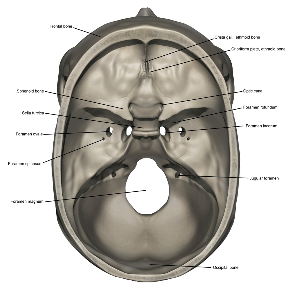
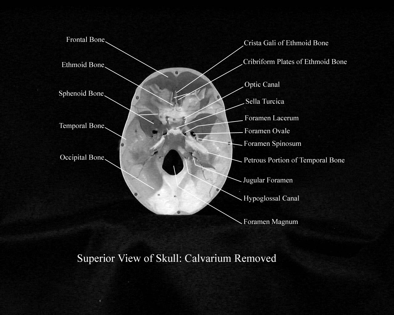
Superior view of the base of the skull Author: Shahab Shahid MBBS • Reviewer: Dimitrios Mytilinaios MD, PhD Last reviewed: October 30, 2023 Reading time: 16 minutes Recommended video: Superior view of base of the skull [21:52] Structures seen from the superior view of the base of the skull. Anterior cranial fossa Fossa cranii anterior 1/3
PPT Chapter 5 The Skeletal System PowerPoint Presentation ID745746
Back to Model Index Page
The Skull · Anatomy and Physiology
1/2 Synonyms: none The human skull consists of 22 bones (or 29, including the inner ear bones and hyoid bone) which are mostly connected together by ossified joints, so called sutures. The skull is divided into the braincase ( neurocr anium) and the facial skeleton ( viscerocranium ).

Superior view of skull bones Skull anatomy, Human anatomy and
1/20 Synonyms: none The posterior and lateral views of the skull show us important bones that maintain the integrity of the skull. The posterior surface protects the region of the brain that contains the occipital lobes and cerebellum .
Skull cranial base, superior view
Synonyms: none In this article we will be focusing on the foramina and fissures located on the inside and floor, or base, of the skull. In a nutshell, a foramen means a hole that can allow various structures to pass through them, ranging from nerves all the way to vessels.

skull labeled anatomy
Synonyms: none The human skull consists of about 22 to 30 single bones which are mostly connected together by ossified joints, so called sutures. The skull is divided into the braincase ( cerebral cranium) and the face ( visceral cranium ). The main task of the skull is the protection of the most important organ in the human body: the brain.
Superior view of human skull anatomy with annotations Poster Print by
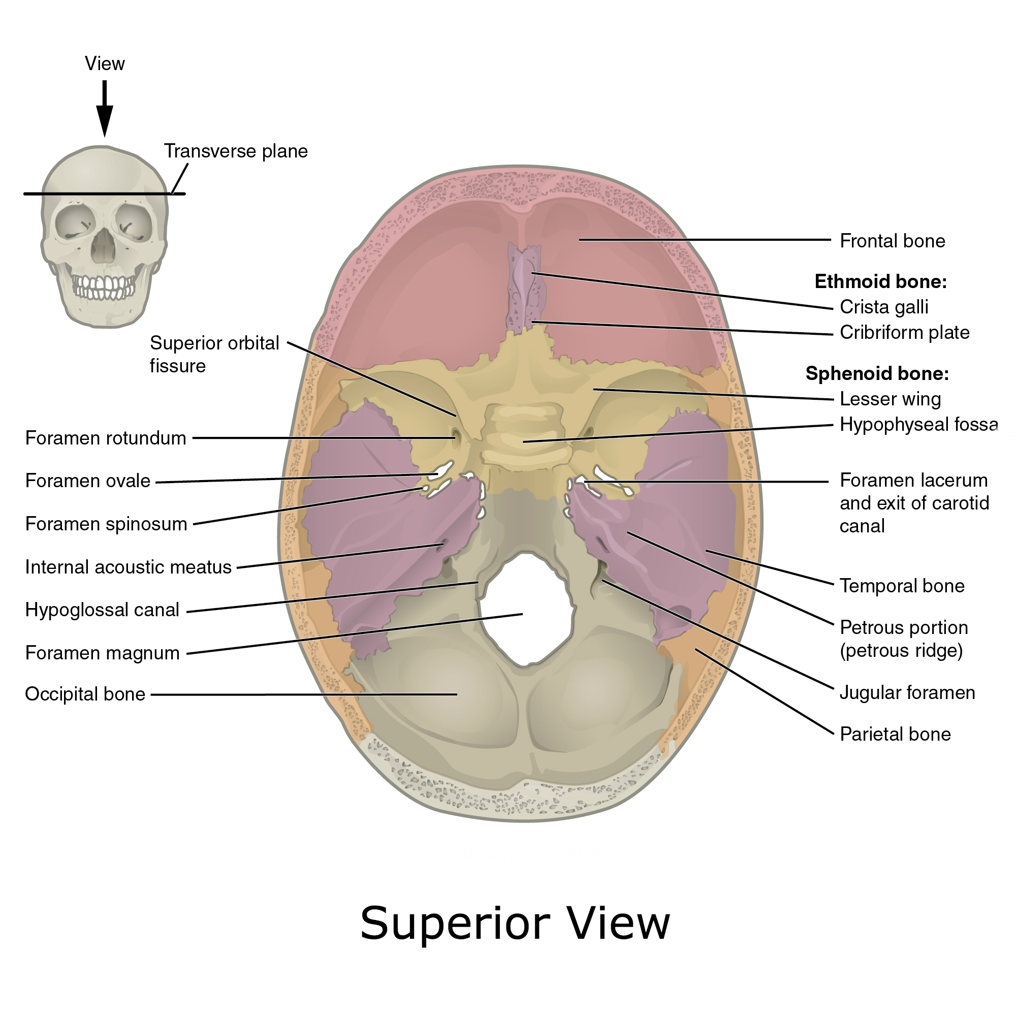
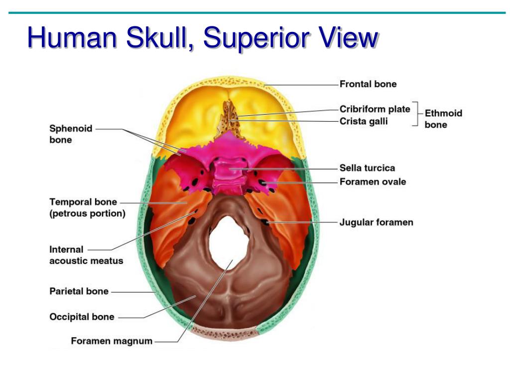
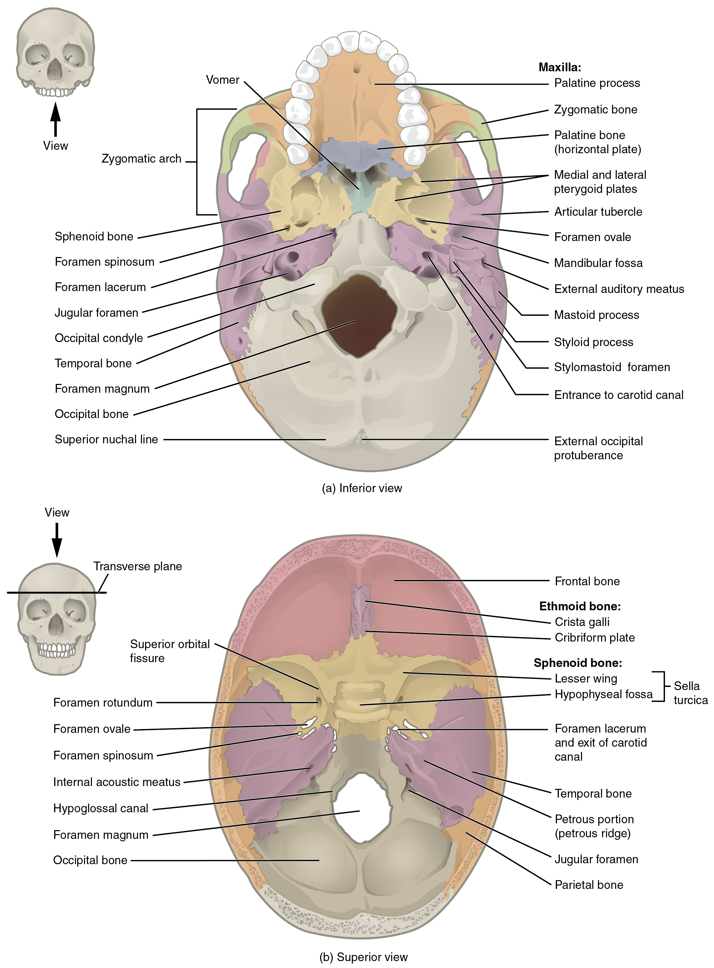
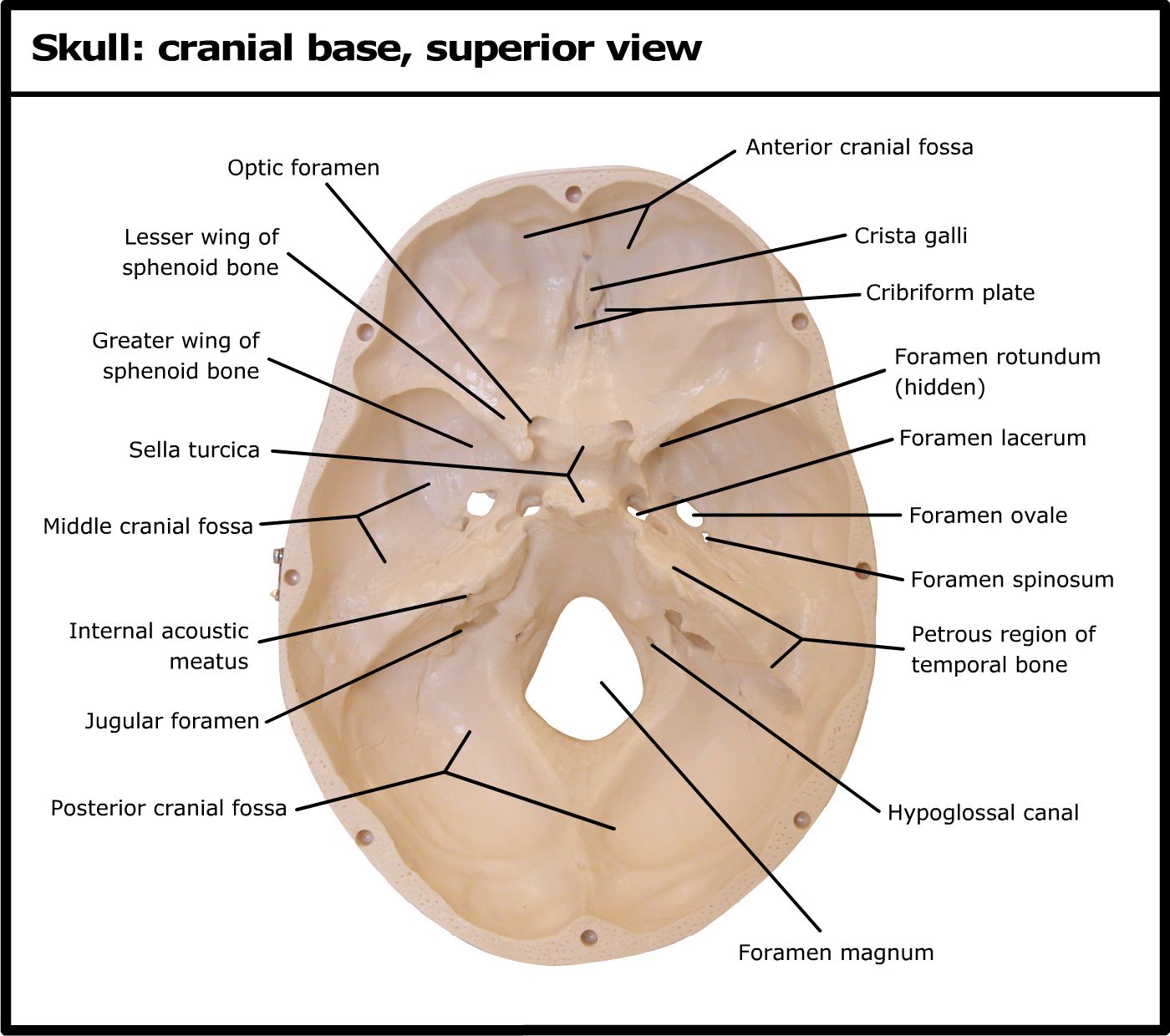
Superior view of the interior of the skull. The skull floor consists of a series of anterior-to-posterior depressions or fossae: anterior, middle, and posterior. The anterior fossa is formed by the orbital plates of the frontal bone, cribriform plate of the ethmoid, and lesser wings of the sphenoid.

Pin on skull anatomy
Tuberculum Sellae. Superior view of the bony skull base. The cranial compartment is separated into three fossae: anterior, middle and posterior. The anterior fossa floor is mostly made up on the frontal bone; in the midline, the ethmoid sinuses are present. The crista galli is the intracranial part of the perpendicular plate of the ethmoid bone.
superiorskullopen1
The cranium (skull) is the skeletal structure of the head that supports the face and protects the brain. It is subdivided into the facial bones and the brain case, or cranial vault ( Figure 7.3 ). The facial bones underlie the facial structures, form the nasal cavity, enclose the eyeballs, and support the teeth of the upper and lower jaws.

Bones of the Head Atlas of Anatomy Anatomía del esqueleto humano
The skull is the skeletal structure of the head that supports the face and protects the brain. It is subdivided into the facial bones and the cranium, or cranial vault ( Figure 7.3.1 ). The facial bones underlie the facial structures, form the nasal cavity, enclose the eyeballs, and support the teeth of the upper and lower jaws.